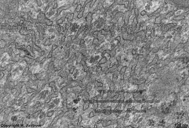
Overview smooth endoplasmic reticulum
(SER):
Pages with explanations are linked to the
text below the images when available
 |
 |
 |
 |
 |
 |
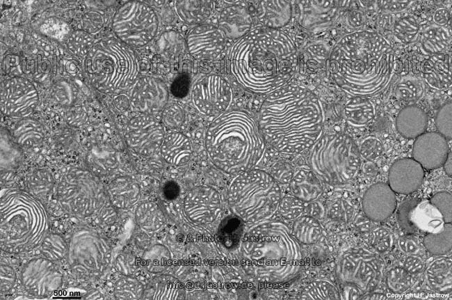
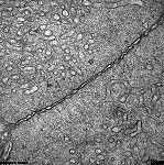
| Detail of SER |
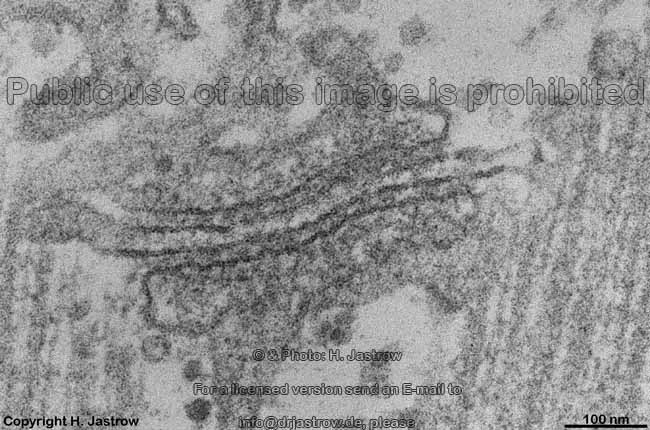
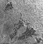
ultra high magnification
of SER (rat) |
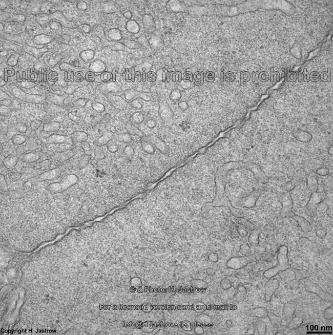
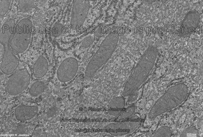
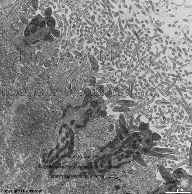
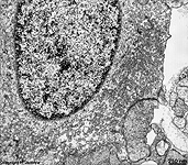
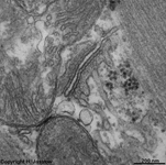
SER in 2 neighbouring
Clara cells (rat) |
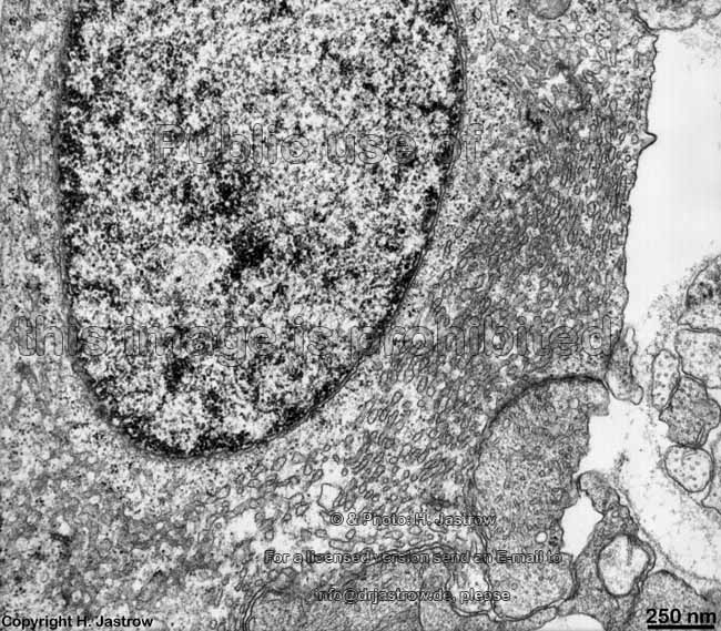
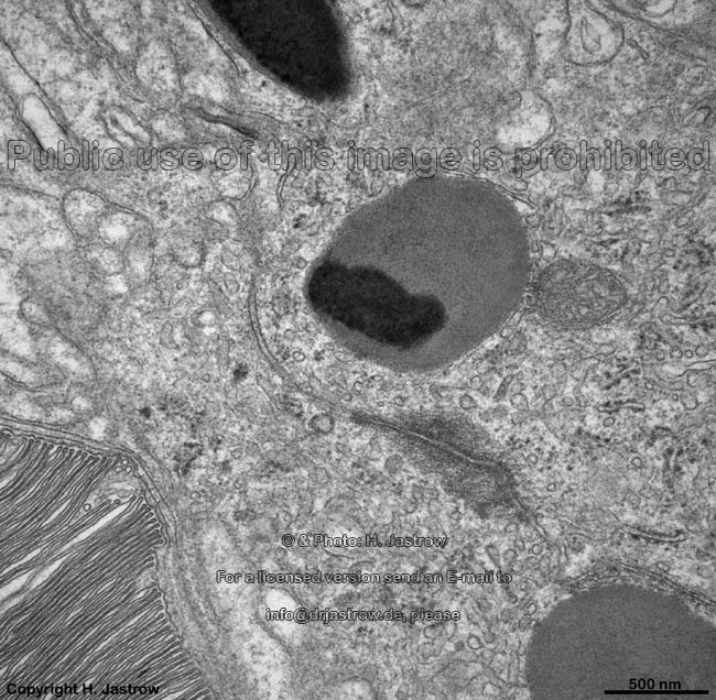
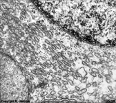
SER in a rat
Pinealocyte |
Detail thereof |
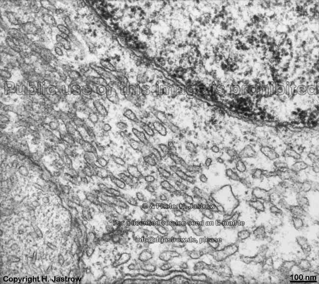
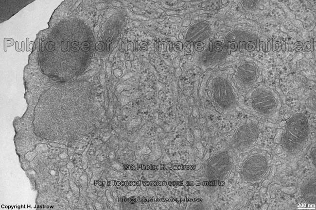
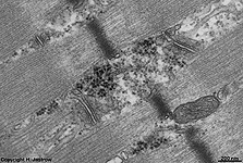
SER and RER
in a liver cell (rat) |
|
|
 |
|
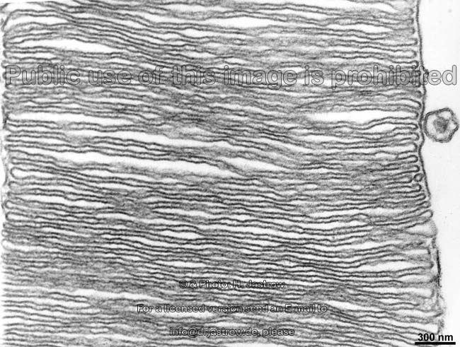
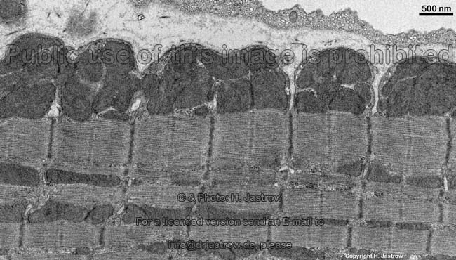
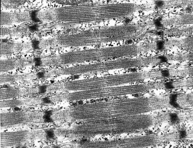
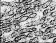
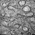
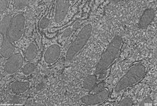
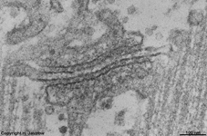
SER as L- Tubuli of skeletal
muscle 1 (Ratte) |
SER as L- Tubuli 2
(Ratte) |
SER as L- Tubuli 3
(Ratte) |
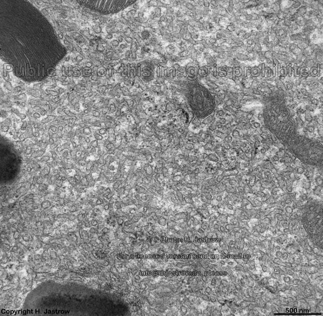
SER in supporting cells,
olfactroy epithelium (rat) |
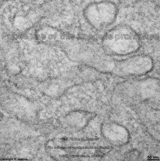
The agranular or smooth endoplasmic reticulum
(SER; Terminologia histologica: Reticulum endoplasmicum nongranulosum)
is a complex three-dimensional network of membranous tubules showing
wider cisterns (Terminologia histologica: Cisternae) as well as tubules
(Terminologia histologica: Tubuli) partly with sac-like protrusions, i.e.
saccules (Terminologia histologica: Sacculi). Its paired membranes
(Terminologia histologica: Membranae) have a distance of 20-50
nm, and in contrast to
RER,
lack
ribosomes. The
outer
surface = cytosolic surface (Terminologia histologica: Facies externa)
borders the cytoplasm while the inner
surface = luminal surface (Terminologia histologica: Facies interna) of
the membranes encloses the lumen (Terminologia
histologica: Lumen). The membranes follow the structural principle
of biological membranes with two electron-dense outer layers enclosing
an inner less-dense lipophilic layer and have a thickness of 6
- 7 nm when cut at right angle. Most cells show much more rough (RER)
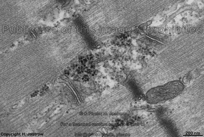
than smooth endoplasmic reticulum. However, a continuity of smooth into
rough ER is not rare and may be seen best in hepatocytes.
This means parts of the membrane system lack ribosomes while other sections
show many of them.
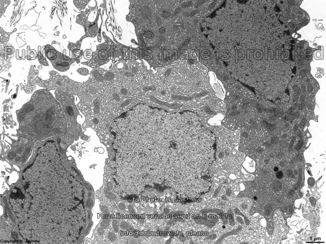
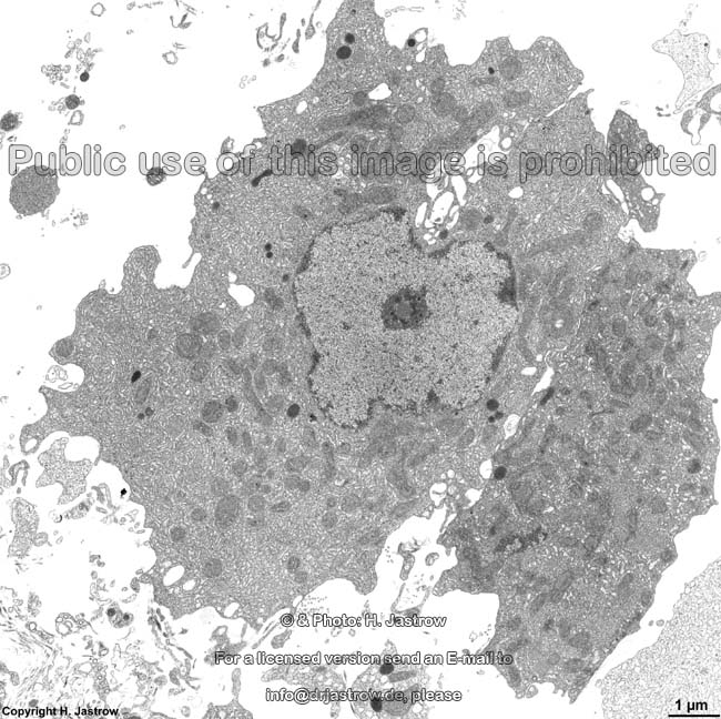
Steroid producing cells possess larger amounts of SER, e.g.
adrenal
gland, testosterone secreting Leydig's
cells of the testis or hormone producing
cells of the ovary [granulosa-
and thekalutein cells]), cholesterin
releasing hepatocytes. Furthermore plenty of
SER is typical for pigment epithelium
of the retina, for Clara-cells
of bronchioles or for supporting
cells of olfactory epithelium.
Also the membrane stacks typical for outer segments of retinal
rods
and cones can be regarded as a special
form of SER. In
skeletal
and heart muscle cells SER forms a network
of thin tubules encompassing each myofibril. Here the SER is called sarcoplasmic
reticulum (Terminologia histologica: Reticulum sarcoplasmicum). Due
to the longitudinal orientation parallel to the myofibrils (photo)
also the term L-tubule has been established for the sarcoplasmic
reticulum.
The SER is similar to the nuclear sheath consisting of an inner and
an outer nuclear membrane which surrounds
the entire nucleus in form of a capsule-like
cistern of the RER showing ribosomes
only on the outer surface
directed towards the cytoplasm. The rare
anulate
lamellas also derive from the SER.
An English page with detailed information and more images is available
in the professional version of this atlas.
--> rough endoplasmic reticulum, cytoplasm,
adrenal
gland, testis,
ovary,
skeletal
muscle,
heart muscle, liver
--> Electron microscopic atlas Overview
--> Homepage of the workshop
Two pictures were kindly provided by Prof. H. Wartenberg,
other images, page & copyright H. Jastrow.