Overview testis (Testis):
Pages with explanations are linked to the
text below the images if available! (Labelling is in German)
 |
 |
 |
 |
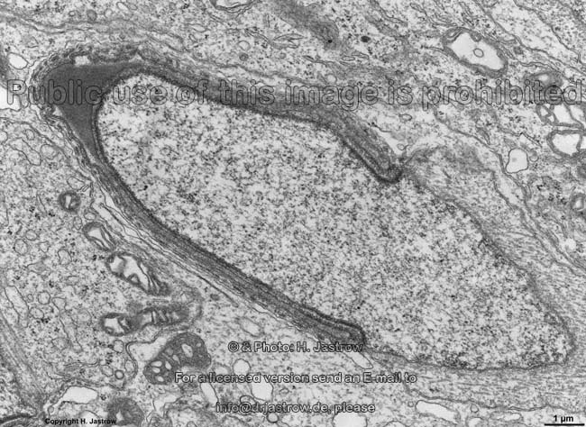
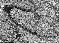
spermatid, acrosome
formation 1 (monkey)
|
spermatid, acrosome
formation 2 (monkey)
|
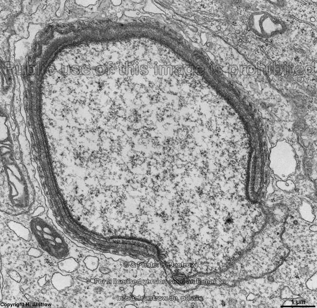
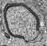
development of acrosomal
cap (rat) |
later stage of acro-
some formation (rat) |
 |
 |
 |
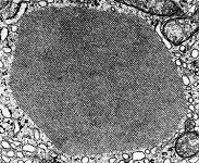 |
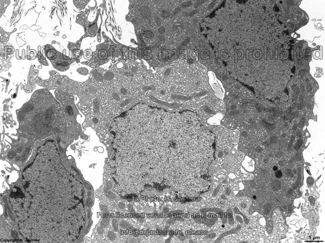
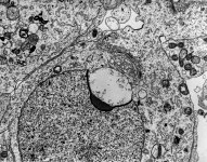
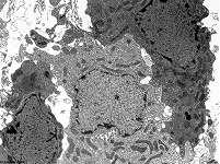
Leydig cells
(rat) |
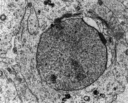
Leydig cells 2 (rat) |
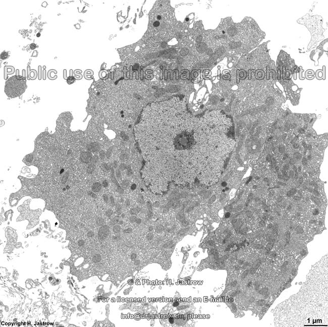
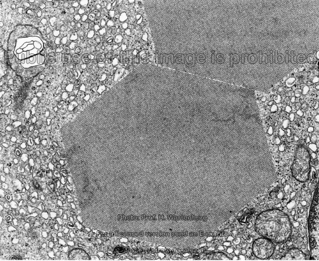
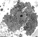
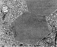
2 Reinke crystals
(monkey)
|
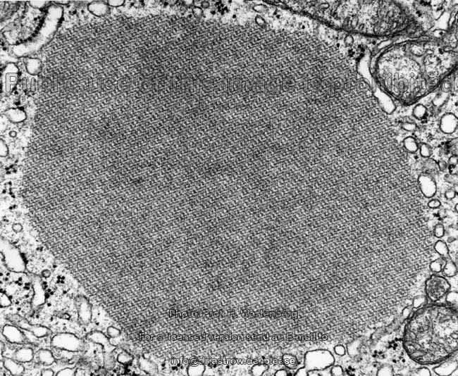
Reinke crystal of a
Leydig cell (monkey) |
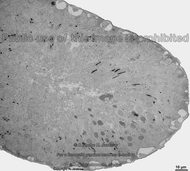
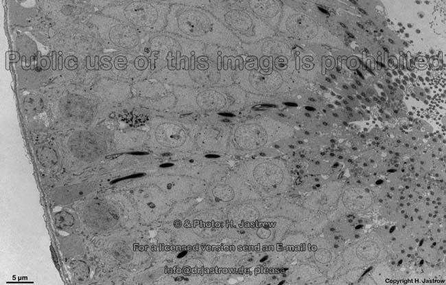
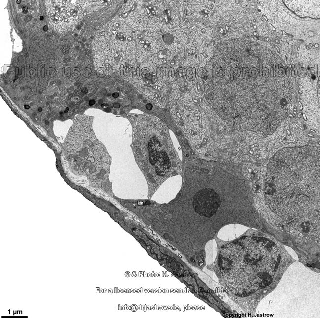
The testis (Terminologia histologica: Testis, Orchis)
contains hundrets of long ducts,
the seminiferous tubules (convoluted seminiferous tubules; Terminologia
histologica: Tubuli seminiferi contorti) in which the maturation and formation
of the male germ cells called spermatozoa takes place. Sertoli
cells (nurse cells, sustentocytes, supporting cells; Terminologia histologica:
Sustentocyti, Epitheliocyti sustentantes) are oriented at right angle to
the basis of the basement membrane and
reach upwards to contact the lumen of the ducts. Different stages of the
maturing germ cells are located in between these nursing cells that
also form by their tight junctions
the blood-testis barrier
(Terminologia histologica: Claustrum haematotesticulare; englisch blood
testis barrier) at the level of the spermatogons to the level of spermatocytes
I.
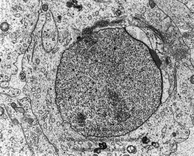
Testosterone producing Leydig's cells (interstitial
endocrine cells; Terminologia histologica: Endocrinocyti interstitiales;
englisch ) are located in the loose connective tissue in between the tubuli
seminiferi. The contain lipid droplets and in some cases may show crystals
of proteins called Reinke crystalloid
(Terminologia histologica: Crystalloidea). Their cytoplasm
contains plenty of tubular-type mitochondria
as well as an abundance of smooth endoplasmic reticulum.
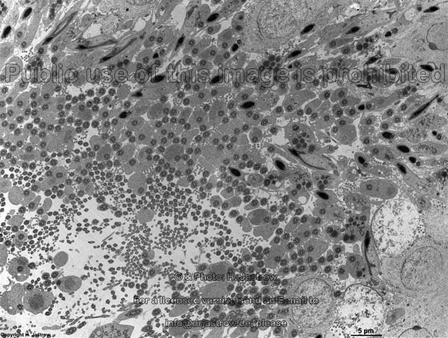
The spermatogenic cells (Terminologia
histologica: Cellulae spermatogenicae) are located at the base of the seminiferous
tubes and during undergoing meiosis give raise to primary
spermatocytes (Terminologia histologica: Spermatocyti primarii) then secondary
spermatocytes (Terminologia histologica: Spermatocyti secundarii)
which then loose great amounts of their cytoplasm
before they become spermatids (Terminologia
histologica: Spermatidia) and finally sperm cells. Sperm
cells (sperms; male gametes; Terminologia histologica: Spermatozoa,
Spermia, Gameti masculini) are the germ cells of males. They only contain
a haploid set of chromosomes in the very
electron-dense nucleus contained in their head
next to a specialised lysosome called acrosome
which is crucial for fertilisation. The body is poor in organelles only
plenty of crista-type mitochondria with
an electron-dense matrix are forming
a sheath around the cilium which is the motile
part of the flagella that extends to the
tail of the sperm.
--> Epididymis, deferent
duct, prostata, epithelium,
crystals,
blood-testis
barrier,
synaptonemal complexes
--> Electron microscopic atlas Overview
--> Homepage of the workshop
Four images were kindly provided by Prof. H. Wartenberg;
other images, page & copyright H. Jastrow.