Overview teeth (Dentes):
electron microscopic images in preparation
|
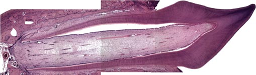
decalcified incisive tooth with periodontal membrane
(histological section, haematoxylin-eosin stain, rat) |
General composition:
Teeth (Terminologia histologica:
dentes) consist of 3 different sustaining tissues, i.e. the enamel,
dentin
and cementum and have a central space (pulp cavity) consisting of
mucoid
connective tissue. From the histological point of view the 32
teeth of the adult are identical to the 20 milk teeth.
The
crown (Terminologia histologica: Corona
dentis) is on top of the tooth neck (tooth cervix; Terminologia
histologica: Cervix dentis) which is covered by the gingiva
(Terminologia histologica: Gingiva) and the
whole structure is anchored by the
root of tooth (Terminologia
histologica: Radix dentis).
Further translation in preparation.
--> clinical anatomy: labelled
image of teeth
--> connective tissue; elastic
cartilage, fibrous cartilage, bone,
elastic
fibres, collagen fibres
--> Electron microscopic atlas Overview
--> Homepage of the workshop
image, page & copyright H. Jastrow.