 |
 |
Clinical Anatomy
in the Internet
Dr.
med. H. Jastrow |
 |

conditions of use! |
| I have made every attempt
to label structures correctly according to the anatomical nomenclature,
however I cannot exclude
mistakes andreject any liability
for eventual errors or incompleteness. Linked pages partly available only
in German
|
Opthalmology
opthalmoscopic image of a normal optic nerve papilla
of the rigt eye
(For higher resolution [1.372 KB] click here,
please !)
upper temporal
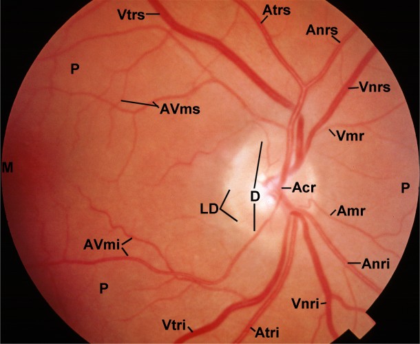 -
lower nasal
-
lower nasal
ACR = Arteria centralis retinae (branching point); Amr
= Arteriola medialis retinae; Anri = Arteriola nasalis retinae inferior;
Anrs = Arteriola nasalis retinae superioris; Atri = Arteriola
temporalis retinae inferioris; Atrs = Arteriola temporalis retinae
superioris;
AVmi = Arteriola et Venola macularis inferior; AVms =
Arteriola et Venola macularis superior;
D = Discus nervi optici (= Papilla nervi optici = optic nerve
papilla = "blind spot"); LD = Limbus disci nervi optici (bordering
wall of the papilla);
M = Macula lutea ("yellow spot" = Fovea centralis; spot of highest
visual acuity on which only cones are present);
P = Pars optica retinae ("viewing part" of the retinal); VCR
= Vena centralis retinae (branching point); Vmr = Venola medialis
retinae;
Vnri = Venola nasalis retinae inferior; Vnrs = Venola
nasalis retinae superioris; Vtri = Venola temporalis retinae inferioris;
Vtrs = Venola temporalis retinae superioris.
--> other optic nerve papilla, overview
entire
retina
--> electron microscopic images of
human retina
--> Clinical Anatomy: Index
--> Homepage of the workshop
The image was kindly provided by Dr. med. O. Schwenn,
university eye clinic Mainz at that time, page & copyright H. Jastrow.