Overview Plasma cells (Plasmocyti):
Pages with explanations (still in German)
are linked to the text below the images when available
 |
 |
 |
 |
 |
 |
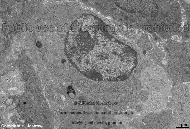
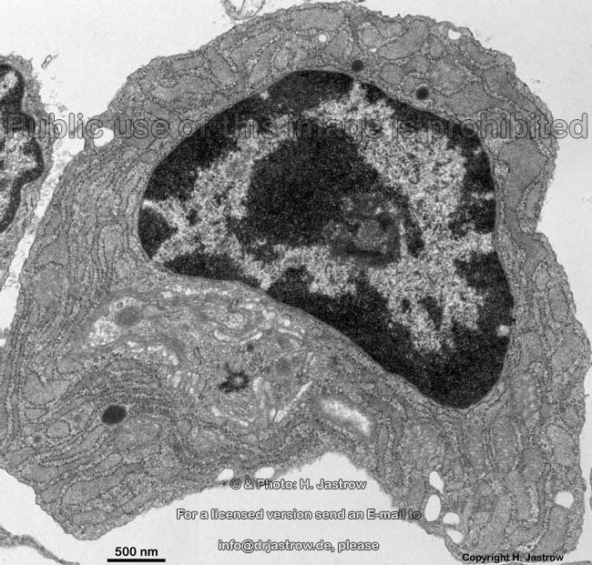
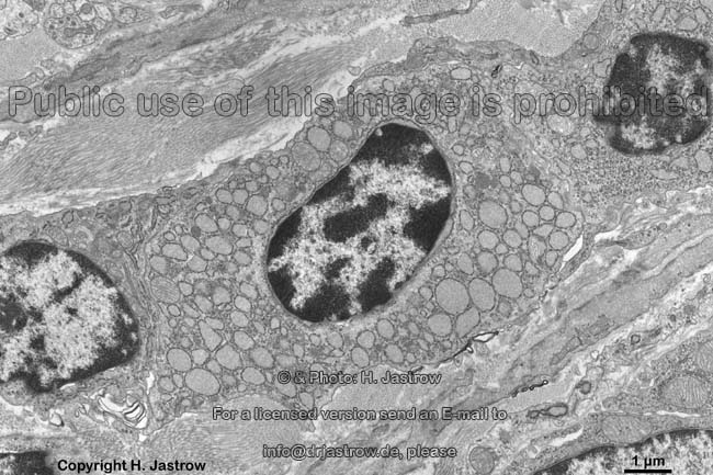
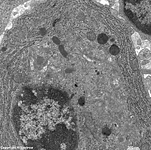
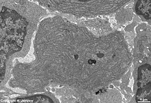
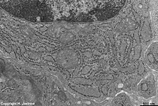
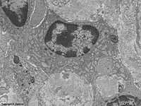
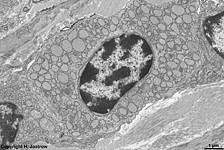
plasma cell 1 from the pharyngeal tonsil
(Tonsilla pharyngea, human) |
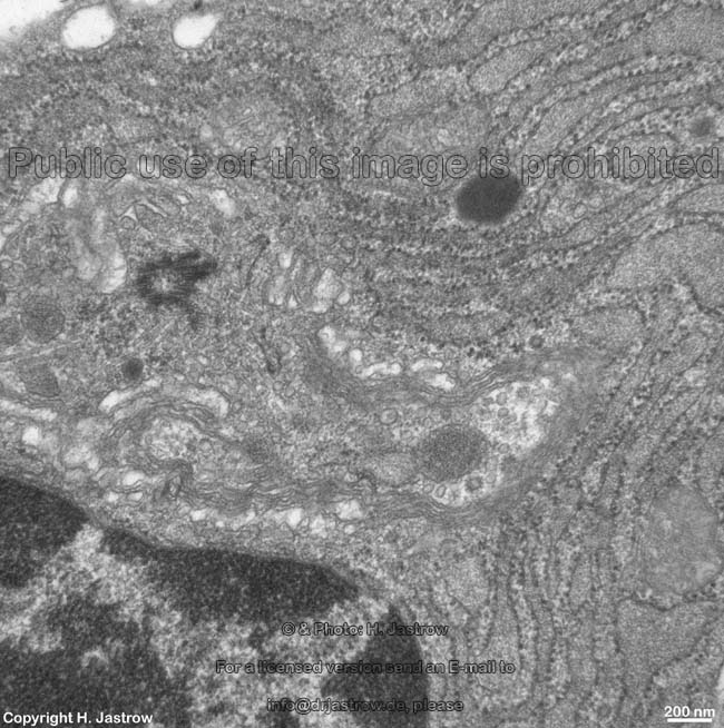
Detail:
cytoplasm |
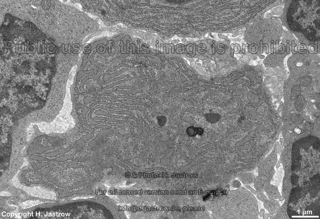
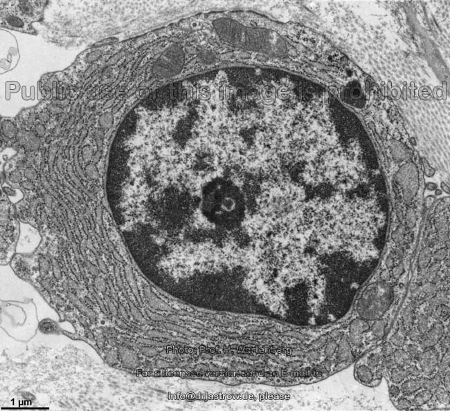
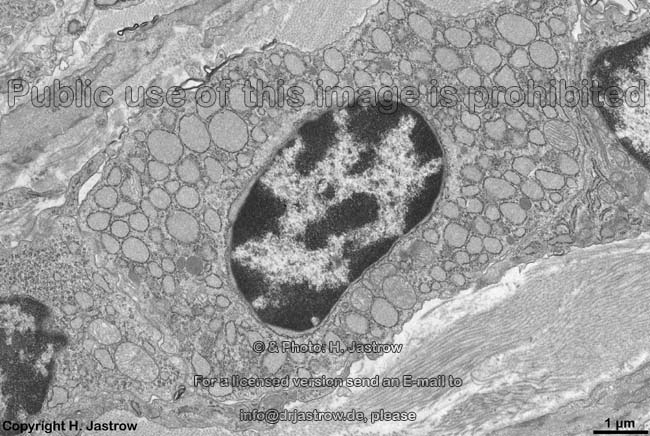
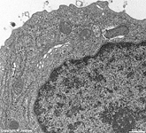
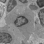
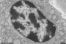
plasma cell 2
Tonsilla pharyngea (human) |
Detail 1:
cytoplasm |
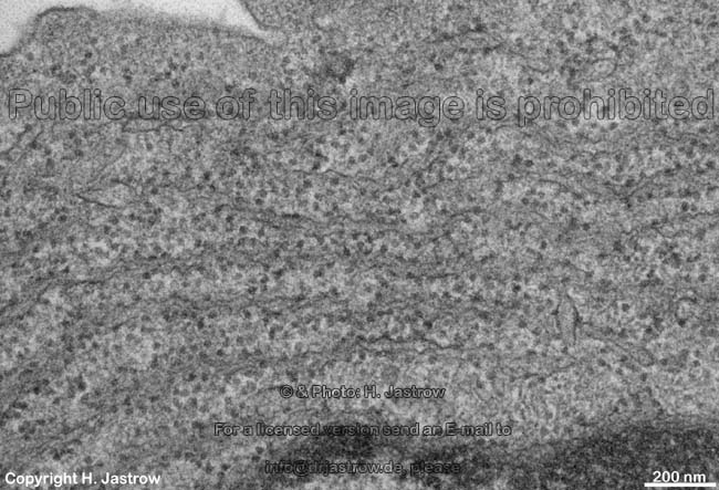
Detail 2:
RER |
Detail 3:
RER |
 |
 |
 |
 |
 |
 |
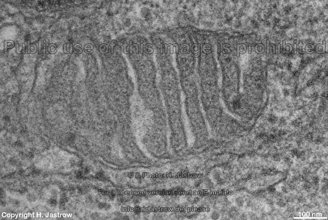
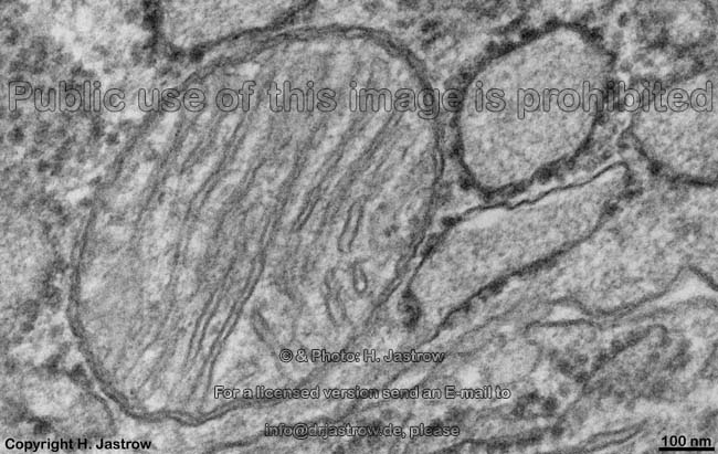
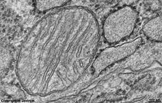
Detail 4:
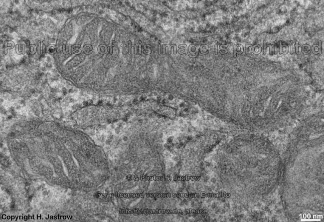
mitochondria (crista-type) |
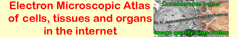
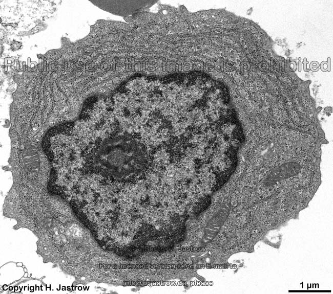
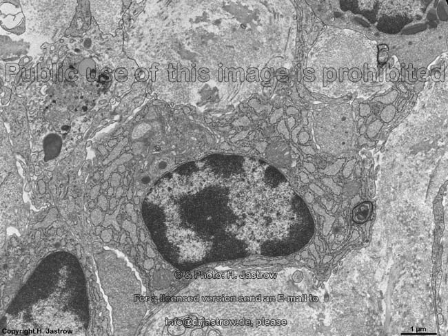
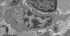
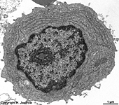
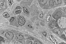
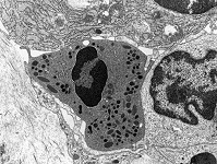
plasma cell 3
Tonsilla pharyngea (human) |
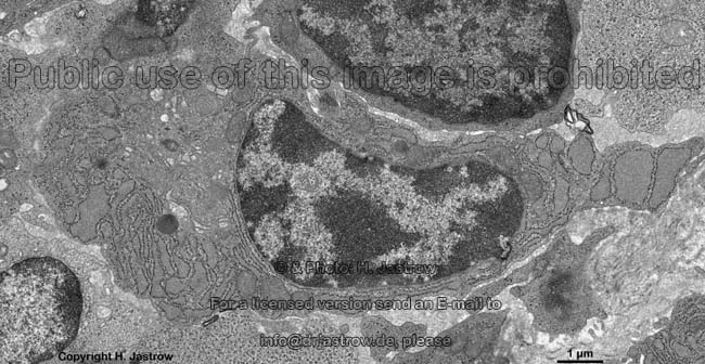
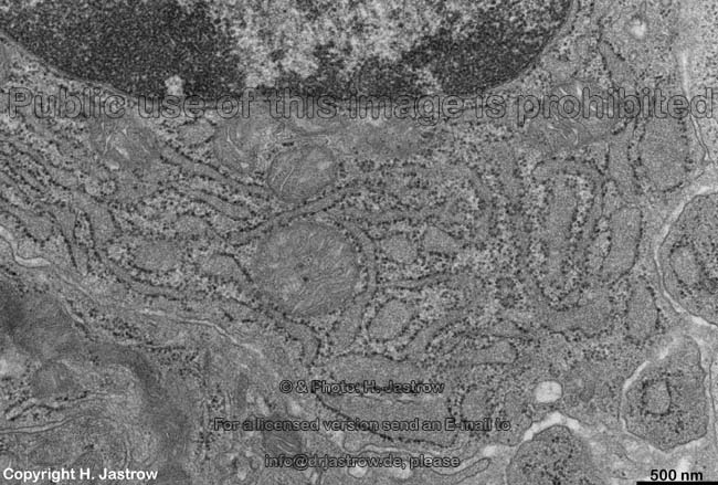
plasma cell 4
T. pharyngea (human) |
detail: nucleus, RER,
Golgi-apparatus |
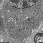
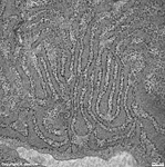
plasma cell 5 from
T. pharyngea (human) |
detail 1:
nucleolus |
 |
 |
 |
 |
 |
 |
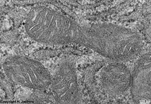
detail 2:
mitochondrium (crista-type) |
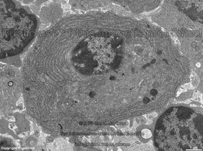
Detail 3: rough
endoplasmic Reticulum (RER) |
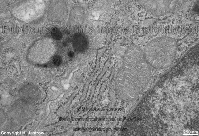
plasma cell 6 from
T. pharyngea (human) |
detail 1:
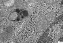
Lipofuscin-vesicle |
detail 2:
RER |
dilated RER plasma cell
T. pharyngea (human) |
 |
 |
 |
 |
 |
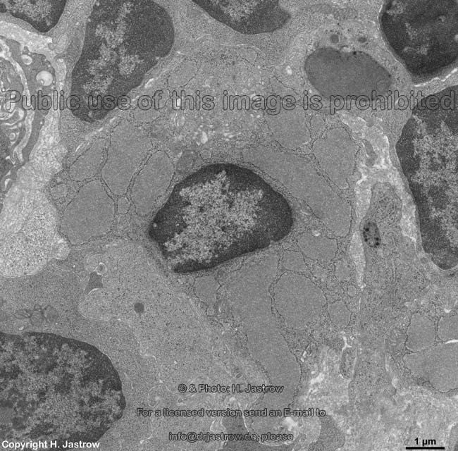
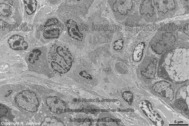
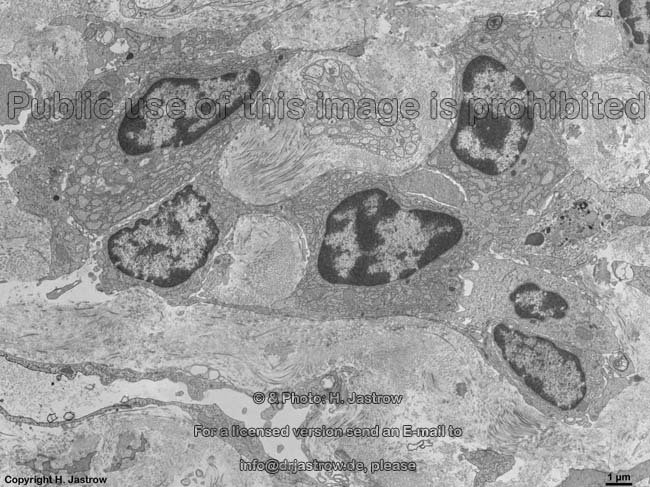
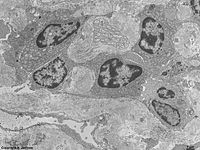
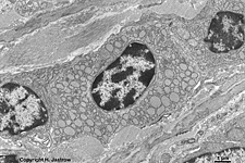
plasma cells of the Lamina propria
mucosae of the colon (Rat) |
4 plasma cells near to a
non-myelinated nerve |
detail thereof:
single plasma cell |
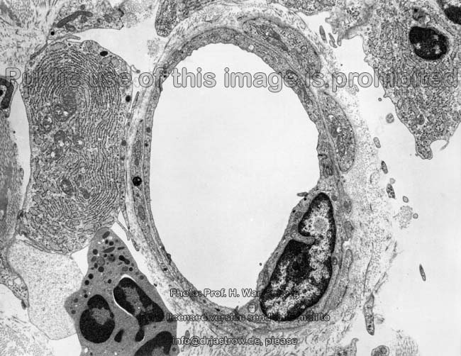
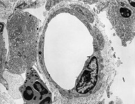
plasma cells close
to a venole (monkey) |
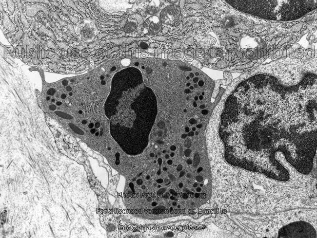
perivascular free connective tissue
cells + a plasma cell (monkey) |
 |
 |
 |
 |
 |
plasma cells of the Lamina propria
mucosae of the colon (rat) |
the next details are
from this plasma cell (rat) |
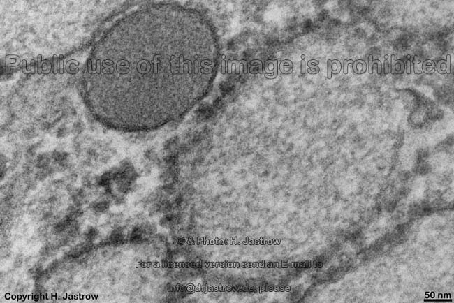
detail: nucleus showing
"spokes of a wheel" structure |
detail: Mitochondrium
of the crista-type |
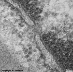
detail:
nuclear pore |
Plasma cells (also called Plasmocytes; Plasmacytes; Terminologia
histologica: Plasmocyti) are free cells of connective
tissue able to move slowly through the latter. Plasma cells are
mature
and active B-lymphocytes synthetising
immunglobulins
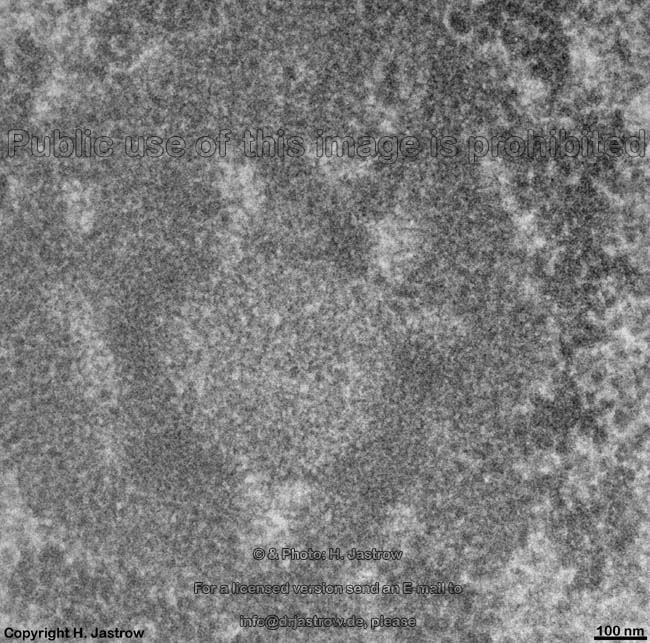
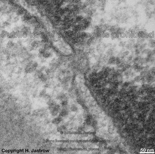
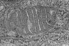
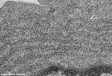
(Ig). Their prominent rough endoplasmic reticulum
(RER) nearly only produces these Ig. Typically it is considerably
widened on several locations. A plasma cell always only releases a single
Ig, i.e. monoklonal antibody, specific for only one epitope of an
antigen. It is most probable that Ig are leaving the RER, diffuse throug
the cytoplasm to be carrierd out of the cell by transmembrane carrier proteins
in the cell membrane since vesicles typical for exocytosis or storage of
secretion products are practically never observable. Further exocytosis
is not detectable and the Golgi-apparatuses
seem too small compared to RER for a fast enough modification of Ig. The
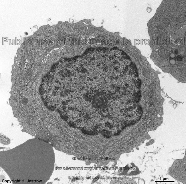
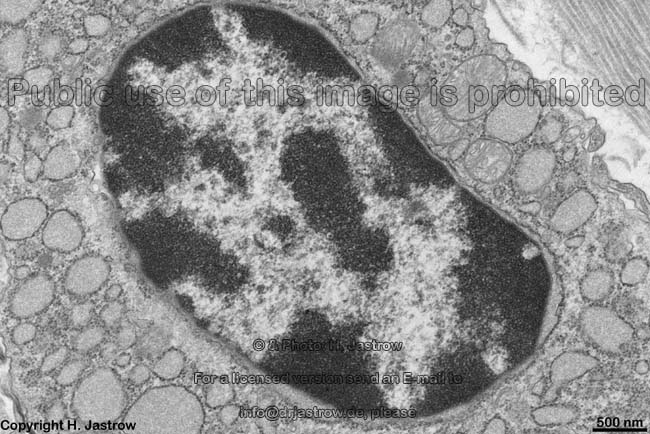
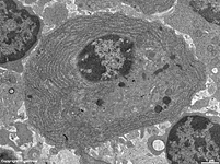
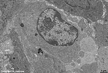
nucleus
shows a typical "spokes of a wheel" structure when centrally cut
with nucleolus associated heterochromatin
in the centre surrounded by a ring of low electron dense euchromatin.
Further nuclear membrane associated heterochromatin
shows wide spaces for a quick penetration of mRNA through the nuclear
pores resulting in low electron dense "spokes of a wheel". Plasma cells
degenerate when not stimulated for several days/weeks. In general plasma
cells are located close to blood vessels.
Some of the synthetised Igs of type E (IgE) stimulate mast
cells and thus support inflammation others directly serve for attack
of antigens (humoral defense).
Depending on the size and arrangements of subunits 5 classes of
antibodies were defined:
IgA relative molecular mass: 162 kDa per monomer;
2 subclasses IgA 1 and 2; secreted by plasma cells in connective
tissue layers located beyond epithelia
thus IgA mainly is present in the mucus
IgD relative molecular mass: 172 kDa; 2 heavy
delta-chains and 2 light kappa or lambda-chains IgD is important
for differentiation of memory and plasma cells.
IgE relative molecular mass: 196 kDa; 2 heavy
eta-chains and 2 light kappa or lambda chains. IgE causes allergic reations.
IgG: relative molecular mass: 150 kDa, ,IgG
passes the blood-placenta
barrier and thus gets into the blood of the embryo / fetus to protect
it before and after birth (inborn immunity).
IgM relative molecular mass: 900 - 935 kDa
in most cases is a very large pentamer consisting of 5, but in some cases
also may be a hexamer consisting of 6 Y-shaped monomers. The latter are
interconnected by a binding protein (J-chain
of 15 kDa).
An English page with much more detailed information and images is only
available in the professional version of
this atlas.
--> blood cells, lymphocytes,
rough
endoplasmic reticulum, bone marrow, connective
tissue, mast cells,
macrophages,
heterochromatin
--> Electron microscopic atlas Overview
--> Homepage of the workshop
Three pictures were kindly provided by Prof. H. Wartenberg;
other images, page & copyright H. Jastrow.