List of abbreviations
Vocabulary
of micros-
copic
anatomy
specialist terms
explained in
English +
German

Every attempt was made to provide correct information and labelling, however any liability for eventual errors or incompleteness is rejected!

dieser Seite

Editor:
Dr. med.
H. Jastrow


Conditions
of use
Overview salivary glands (Glandulae salivariae):
Pages with explanations are linked to the text below the images if available! (Labelling is in German)
Gl. submandibularis
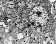
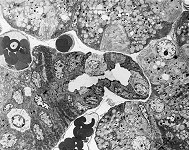
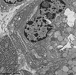
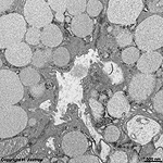
serous acinar cell (monkey)
acinus + secretory
vesicles 1 (monkey)
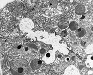
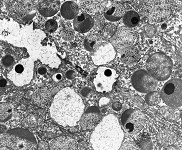
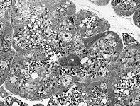
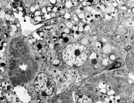
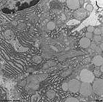
acinus + secretory
vesicles 2 (monkey)
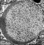
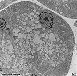
serous secretory
vesicle (monkey)
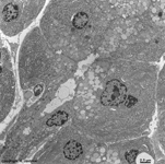
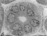
acini and striated duct
(monkey)
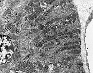
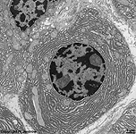
striated duct
detail (monkey)
Glandula submandi-
bularis (monkey)
Gl. submandibularis
serous acinus (monkey)
mucous + serous secretory
vesicles sublingual gland (rat)
mucous epithelial cell of the
sublingual gland (rat)
detail: mucous
 vesicles (rat)
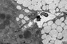
Ohrspeicheldrüse 1
(Ratte)
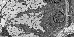
Ohrspeicheldrüse 2
(Ratte)
Detail: RER
+ Vesikel (Ratte)
Ohrspeicheldrüse 3
(Ratte)
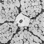
Parotis: Azinus + 
Schaltstück (Ratte)
Azinus Ohrspeichel-
drüse (Ratte)
Schaltstück Ohr-
speicheldrüse (Ratte)

There are three major salivary glands: parotid gland (Glandula parotis), submandibular gland (Glandula submandibularis) and the sublingual gland (Glandula sublingualis) in the mouth. Further, there is the pancreas (Pankreas) that delivers its secretions into the lumen of the duodenum. Further small salivary glands are located in the tongue and in the mucosa of the palate.
Oral salivary glands are all eccrine exocrine glands and are classified as:
name of the group name of the gland secretion mode histological characteristics position duct opening
Glandulae salivatores
majores
main salivary glands
Gl. parotidea
parotid gland
serous narrow acini, intercalated ducts, long striated ducts,
fat cells in the stroma; one large excretory duct
in the retromandibular fossa Papilla ductus parotidei
opposite to molars 16,17 / 26,27
Gl. submandibularis
submandibular gland
seromucous
(serous>mucous)
tubulo-acinar end pieces, v.Ebner demilunes, myoepithelial cells,
striated ducts, one large excretory duct
in the submandibular space Caruncula sublingualis on the
base of the frenulum linguae
Gl. sublingualis
sublingual gland
mucoserous
(mucous>serous)
tubulo-acinar end pieces, v.Ebner demilunes, myoepithelial cells,
short striated ducts, many excretory ducts
lateral posterior above
the mylohyoid muscle
several openings on top of tips
along the sublingual plica
Glandulae salivatores
minores
small salivary glands
Gll. sublinguales minores
small sublingual glands
mucoserous
(mucous>>serous)
hardly any acini, mainly tubular end pieces, no striated ducts,
The posterior end of this group is called the glossopalatine glands
along the sublingual plica
on the floor of the mouth
several openings on top of tips
along the sublingual plica
Gll. labiales
labial glands
seromucous
(serous>mucous)
compound tubulo-acinar, no striated ducts in the mucosa of the
upper and lower lip
mucosa of the upper
and lower lip
Gll. buccales
inner buccal glands
mucoserous
(mucous>serous)
compound tubulo-acinar, no striated ducts in the mucosa of the cheek several openings into the
mucosa of the cheek
Gll. molares
outer buccal glands
mucoserous
(mucous>serous)
compound tubulo-acinar, no striated ducts outside on top of the
buccinator muscle
several openings into the
mucosa of the cheek
Gll. palatinae
palatine glands
mucous compound tubular, no striated ducts in the palatine mucosa several openings into the
mucosa of the palatum
Glandulae linguales
lingual glands
Gl. lingualis anterior (Nuhn)
anterior lingual gland
mucous compound tubular, no striated ducts beneath the tip
of the tongue
mucosa beyond the tip
of the tongue
Gll. linguales posteriores
posterior lingual glands
mucous compound tubular, no striated ducts beyond the lingual tonsil crypts of the lingual tonsil
Gll. gustatoriae
von Ebner's glands
serous branched acinar glands  mucosa beneath tongue
papillas with taste buds
open into the ditches
between lingual papillas
construction of a larger salivary gland:
The major part of the secretion is produced by the epithelial cells around the acini of the gland. After the acinar or tubulo-acinar end piece (Terminologia histologica: Portio terminalis) which may also be a demilune of Ebner (Terminologia histologica: Semiluna serosa) small intercalated ducts (Terminologia histologica: Ducti intercalati) with a monolayer of cubic epithelium transport the secretion into an intralobular duct whose monolayered columnar epithelial cells show a "basal striping". This striping is caused by regularly stacked mitochondria in deep infoldings of the cell membrane (--> example image) and is visible in light- and electron microscopy. The pancreas (and the lacrimal gland), however, are devoid of this "basal striping". The corresponding parts of their excretory ducts are just called intralobular ducts. When such a duct leaves the lobe of the gland, it becomes an interlobular duct (Terminologia histologica: Ductus interlobularis) and is surrounded by connective tissue. The columnar epithelium then begins to show an additional basal cell layer, i.e. is a bilayered columnar epithelium. Depending on the size of the gland many of such ducts join to form one or more interlobar ducts (Terminologia histologica: Ducti interlobares) which further join into a main excretory duct which has a pseudostratified epithelium.

--> exocrine merocrine glands, glands, epithelium, secretory vesicles
--> Electron microscopic atlas Overview
--> Homepage of the workshop


Eight images were kindly provided by Prof. H. Wartenberg; other images, page & copyright H. Jastrow.