Overview synapse (Synapsis):
Pages with explanations are linked to the
text below the images if available! (Labelling is in German)
 |
 |
 |
 |
 |
 |
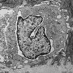
synapse on a Merkel
cell, skin (rat) |
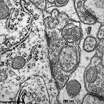
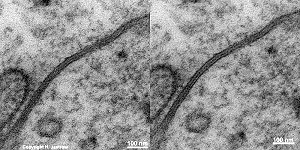
type I synapse
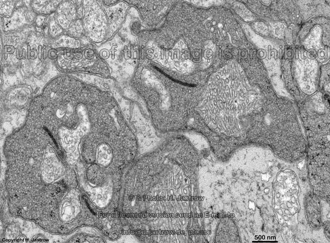
Cortex cerebri 5 (rat) |
synapses in cerebral
cortex (rat) |
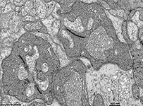
ribbon synapses of the
retina (rat) |
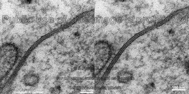
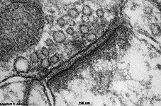
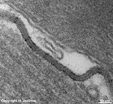
electrical synapse = gap junction = nexus
stereo image, pilar cell, inner ear (rat) |
nexus, intercalated
disk, heart (rat) |
A general distinction is made between electrical and chemical synapses.
Electrical synapses
(non-vesicular synapses; Terminologia histologica: Synapses nonvesiculares;
Synapses electricae) correspond to nexus
(gap junctions; maculae communicantes; Terminologia histologica: Maculae
communicantes; Nexus) and consist of tunnel
proteins (further details here). They provide
faster
signal transduction than chemical synapses and by
a process called electrotonic coupling generate an ultrafast balance
of membrane potentials of coupled neurons. This allows a synchronized conduction
of impulses in strands of cells coupled by nexus which is of importance
e.g., during maturation of the brain in the foetal period. In adults, however,
nexus are present in relevant number only in some few areas still: they
synchronize inhibition of Purkinje's
cells by coupling basket
and stellate cells of the cerebellum;
couple glial cells of the CNS,
olfactory bulb (Bulbus olfactorius), sensory cortex of the Gyrus postcentralis
(main sensory area for body perception). Nexus
are bidirectional i.e., the signal transduction caused by direct
flow of ions may run in both directions. Nexus are even present
between epithelial cells of glands in order
to synchronise their action.
mixed synapses (missing in Terminologia
histologica: Synapses mixtae) partly show connexons
typical for electrical synapses and directly adjacent chemical synapses.
They are very rare and shall be restricted to few nuclei of brain nerves.
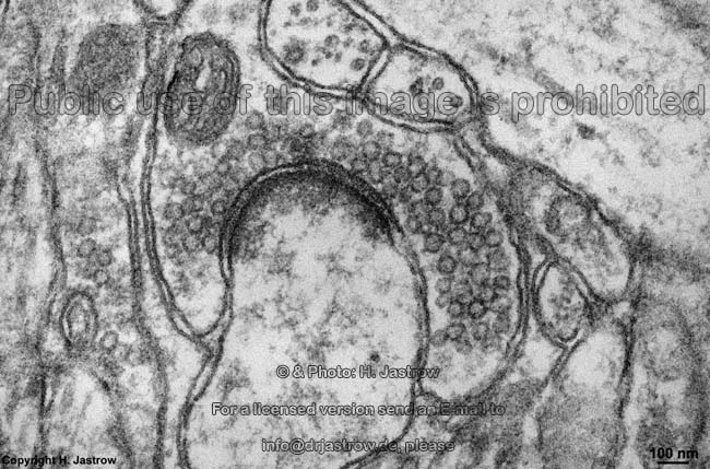
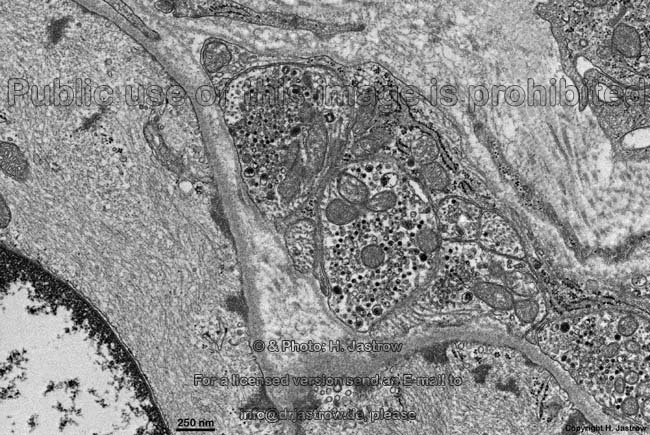
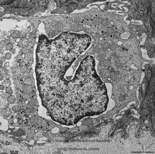
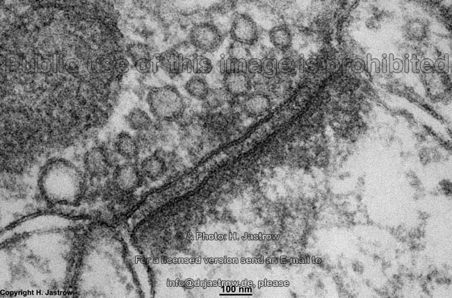
Chemical
synapses (vesicular synapses; Terminologia histologica: Synapses
vesiculares; Synapses chemicae) are typical for the central (brain
and spinal chord) and the peripherical nervous system (all other kinds
of nerve tissue). In spot-like nearly
round areas with diameters of a few hundred nanometres chemical synapses
in most cases are asymmetrical, i.e. pre-
and
postsynaptic membranes
have different features causing a unidirectional conduction of impulses.
Types of chemical synapses are descired below.
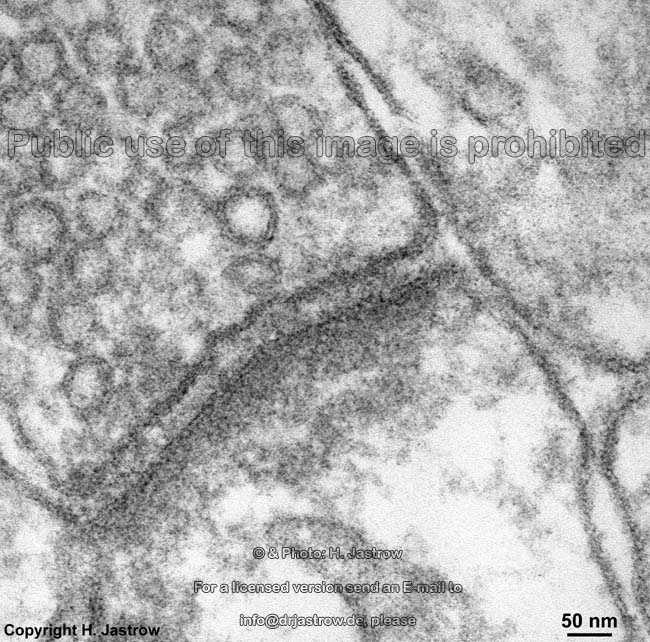
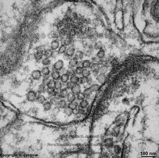
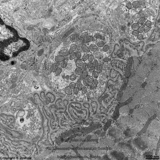
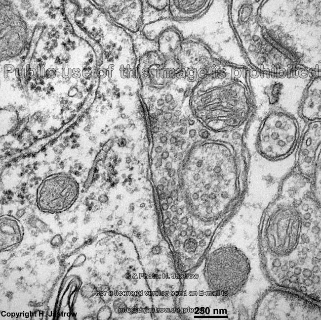
Presynaptic region (Terminologia
histologica: Pars presynaptica)
The presynaptic side shows a widening of the
terminating nerve cell process called axon
(Terminologia histologica: Axon), which
is termed terminal bouton (Terminologia histologica: Bulbulus terminalis). In
many instances axons split into
several axon collaterals (Terminologia histologica: Rami collaterales
axonis) just shortly before their end which then form such boutons. Terminal
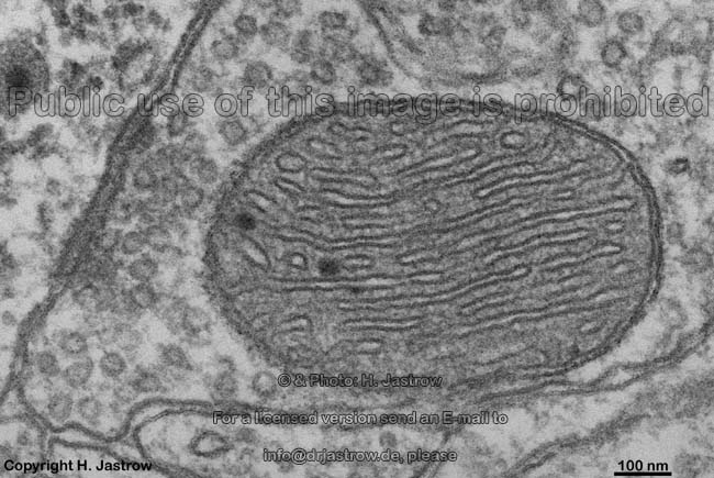
boutons contain plenty of crista-type mitochondria
providing energy by their ATP synthesis, further
actin
filaments (called neurofilaments
here) as well as microtubules
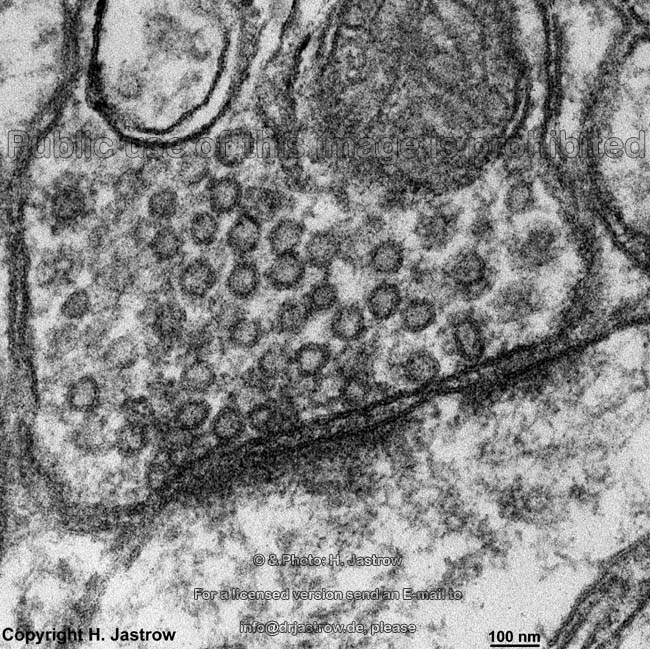
(termed neurotubules here) and some hundred to thousands of neurotransmitter
vesicles (Terminologia histologica: Vesiculae synapticae; Vesiculae
presynapticae: diameter: 40 - 60 nm). From point of view of function the
latter are the most important component of the boutons. Synaptic
vesicle linking strands (Terminologia histologica: Vincula vesicularum
synapticorum) are 30 - 60 nm long, few nanometres thick, slightly electron-dense
filamentous structures keeping the vesicles close to the cell
membrane. They contain synapsin,
a fibrous phosphoprotein and link the synaptic vesicles to actin-
and spectrin filaments of the
cytoskeleton.
Further synapsin is the morphological correlate for fine filamentous interconnections
of the vesicles to each other. It comprises ~6 % of all membrane proteins
of these vesicles. Synapsin is the substrate of cAMP-dependent- and calcium-calmodulin-dependent
proteinkinases being phosphorylated with raising level of calcium ions
(Ca++) in the cytoplasm. This
is required for disconnection of the vesicles from the cytoskeleton
a prerequisite for their release.
An English page with much more detailed information and further images
is available in the professional version
of this atlas.
--> electrical synapse, synaptic
bodies, nerve tissue, nerve
--> sensory organs, nerve
fibres, nerve terminals,
central
nervous system, retina
--> Electron microscopic atlas Overview
--> Homepage of the workshop
Images, page & copyright H. Jastrow.