Overview pseudopods (Pseudopodia):
Pages with explanations are linked to the
text below the images if available! (Labelling is in German)
 |
 |
 |
 |
 |
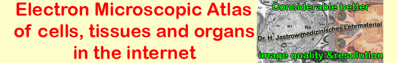
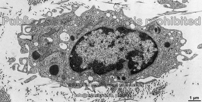
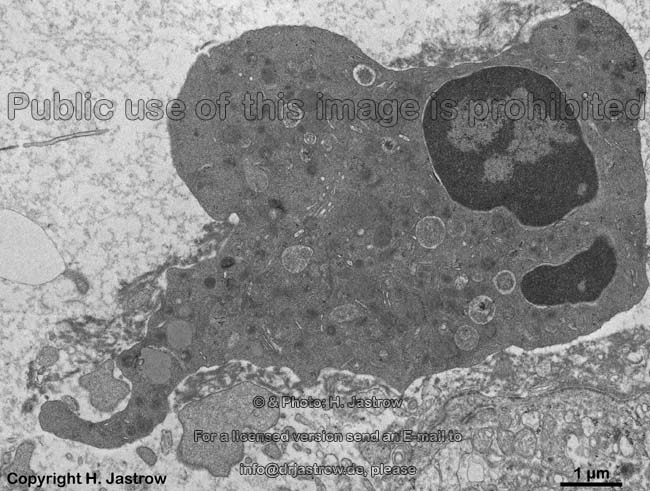
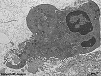
human eosinophilic gra-
nulocyte with pseudopods |
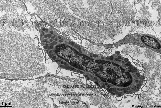
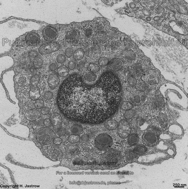
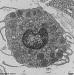
similar human eosino-
philic granulocyte |
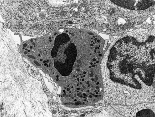
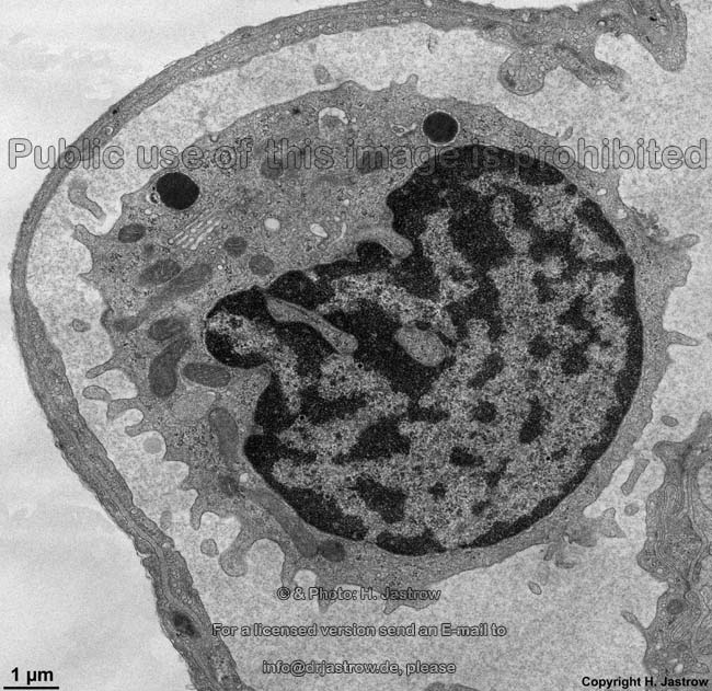
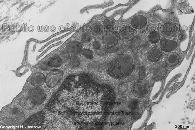
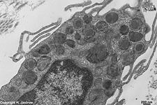
human neutrophil protrudes its
pseudopodfor phagocytosis |
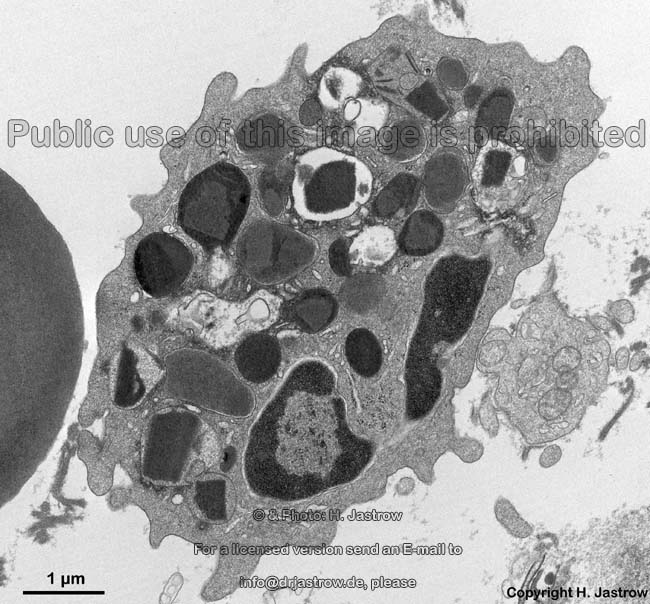
human mast cell with
thin pseudopods 1 |
human mast cell with
thin pseudopods 2 |
Pseudopods (Terminologia histologica: Pseudopodia) are
foot-like elongate thin mobile processes of cells containing
cytoplasm
and actin filaments. They are
no straight protrusions but much more irregularly formed than microvilli.
Pseudopods may arise from cytoplasmwithinfew
minutes and may also be retracted in similar time. Pseudopods
are actively mobile since they contain actin
as well as myosin filaments.
Due to oriented polymerisation and depolymerisation of actin
filaments in the anterior part of a pseudopod a short-time adhaesion
of the cell membrane to extracellular
structures and contraction of the posterior part of the cytoplasm
in case of disattachment in the posterior part of established contact points
of the cell to surrounding structures the whole cell is drawn forward by
the pseudopod. Hereby myosin 1 binds
the cytoskeleton to actin
filaments which polymerise anteriorly, i.e. which further grow in this
direction. In the intermediate part contractile stressfibres
(Terminologia histologica: Fibrae tensionis) consisting of
actin
filaments and interposed myosin 2
bind to cell-matrix contacts. Contraction of the stress fibres then together
with alterations of the cytoskeleton
result in a shortening of the cell which anteriorly as mentioned is attached
to adjacent matrix via outgrowing pseudopods which altogether results
in anterograde movement. Thus pseudopods allow the cell to move actively
whereby the velocity of this movement is influenced by temperature,
local concentration of ions and pH value changes as well as by chemotactic
substances. Neutrophilic granulocytes can
migrate with a speed of 20 to maximal 60 µm in a minute.
Occurrence:
The following cells are able to migrate via pseudopods: free
cells in connective tissues:
plasma cells,
macrophages,
eosinophilic
granulocytes, mast cells; blood
cells: all kinds of granulocytes,
lymphocytes,
monocytes.
Further, pseudopods are important for phagocytosis
of macrophages: pseudopods protrude in
direction of a foreign body, e.g. a bactrium, reach it, embrass it and
finally by contraction draw it into the cell while the pseudopods fuse
with each other before the phagocytosis is finished.
--> microvilli, kinocilia,
stereovilli,
cell
surface structures, cell membrane
--> Electron microscopic atlas Overview
--> Homepage of the workshop
Dr. E. Schiller, Prof. H. Wartenberg and HSD Dr. Klinger
kindly provided one image each; other images, page & copyright H. Jastrow.