transverse sections of the Visible Human male
Conditions
of use
| Homo sapiens dissecatus
transverse sections of the Visible Human male |
Conditions of use |
|||
| Editor: Dr. H. Jastrow | ||||
1. Pages with original
sections, CT & MRI
2. Pages with labelled
sections
3. Pages with overviews
of sections
4. Index page
5. Pages providing motion
pictures
6. Page with X-ray images
general information:
To use this atlas you need a web
browser. Pages are optimized for use with a screen resolution of 1,600
x 1,200 pixels.
The navigation is managed by symbols
in order to be independent of language. It is explained on the linked pages
(1-5). Inevitable annotations are in Latin or English.
The digitized cross-sections base
on high-resolution rescans of the original films taken from the visible
human male sections. One pixel of these 24-Bits of colour images represents
an area of 0.14 x 0.14 mm of an original section and the distance between
the sections is 1 mm. The compressed RAW image data files were kindly provided
by the National Library of Medicine (U.S.A.) as well as the corresponding
16-Bits of grey Computer tomography (CT) and nuclear magnetic resonance
tomography images (MRI). Original MRI in the orientation of the sections
(transverse) were provided only for head & neck. MRI in other regions
were calculated from data provided in the frontal plane.
All images were trimmed and are
provided as optimised 24-Bits of colour compressed JPEG files (maximum
quality = professional-, high quality = student- and low quality = www-version
of the atlas).
All original sections and CT/MRI
were cleaned from artefacts as far as possible. Missing sections and parts
of structures destroyed by sectioning were carefully reconstructed.
All available CT/MRI are shown
in original resolution next to the sections with best correspondence. Pages
with digitized original sections provide them in a size adapted to the
screen in order to allow viewing of the radiological images as well. By
clicking the 100% symbol the digital sections are shown as graphics in
the resolution in which they are offered. It depends on the version of
the atlas: original resolution - professional version, 60% - student version,
30% - www version. This also applies to the labelled sections.
Every attempt was made to
label as many structures as possible and reasonable
according
to the anatomical nomenclature (Terminologia anatomica)
and
to provide correct information, however any liability for eventual errors
or incompleteness is rejected. Labelling is
being continued for all sections. If you should encounter any imperfections
notify me via
E-mail, please.
|
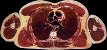
of the sections agrees to the standard established in Radiology, i.e. you look at the sections from below. See example on the right! |
|
Look up the used terms in the
(click the icon!) |
Use of this atlas is
subject to the linked Conditions |
motion pictures through all sections of the Visible Human male, CT- and MRI click here! |